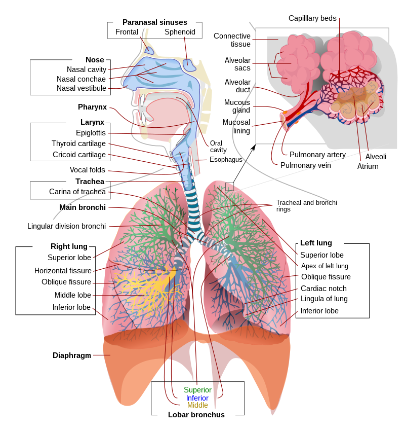
Video Assisted Thoracoscopic Surgery is a minimally invasive chest surgery where a scope is inserted through small incisions. The technique is used to perform a variety of different procedures including esophagus and lung surgery and biopsies to diagnose lung cancers.
What is it?
As with many minimally invasive surgeries, VATS is used instead of open surgery because it generally causes less pain, results in fewer complications, and shortens recovery time. Using a special instrument called a thoracoscope, the surgeon will be able to access the chest and lungs, transmitting images inside the body to a video monitor. This thoracoscope, along with other surgical instruments, will allow the doctors to operate on the patient without making any large incisions. VATS can be used to treat many different medical conditions and can be used for both diagnostic and therapeutic surgery. Some common conditions treated with VATS include esophagus surgery, lung surgery, biopsies, gland or lymph node removal, hernia repair, and fluid accumulation in the chest.
What should I do to prepare?
Exposure to second-hand smoke should be limited and, if possible, prevented at least two weeks prior to surgery. However, it would be best to prevent exposure months before. You will also be asked to stop taking blood thinners and other blood medications. You may be asked to perform several breathing and aerobic exercises to strengthen your cardiovascular system in the weeks before the surgery. As with other surgeries, eat foods recommended by your doctor the day before and do not eat/drink anything past midnight the night before your surgery.
What happens during the process?
Patients will generally be given general anesthesia for VATS procedures, but they may also receive an epidural for more pain relief. A catheter may also be inserted into the patient’s bladder seeing as the epidural may make it difficult to urinate. The patient may be lying on their back or side depending on the position the surgeon requires to perform the surgery. After making a few small incisions, the surgeon will insert the thoracoscope and other operating instruments. The surgeon will then operate on the patient using the images sent by the thoracoscopic camera. After the procedure is complete, the tools are removed and one or more tubes will be left in the chest. These will drain the excess air and fluids from the chest or abdomen. After this, the surgical incisions are closed and bandaged.
What are the risks and potential complications?
Some associated risks and complications include, but are not limited to, excessive bleeding and infection, fluid leaks from the chest or lungs, chest pain, shortness of breath, anesthetic complications, and risk of conversion to an open surgery
Disclaimer:
All GlobeHealer Site content, including graphics, images, logos, and text, among other materials on the site are for educational purposes only. This content is not intended to be a substitute for professional medical advice, and you should always contact your physician or qualified health provider for information regarding your health. Information on this site regarding the overview, diagnosis, and treatment of any kind should be looked at, in addition to the advice and information of your health care professional. Do not disregard medical advice or delay seeking treatment or medical advice due to information found on the GlobeHealer site.
If there is even the possibility that you may have a medical emergency, seek treatment, call your doctor, or call your local emergency telephone number immediately. GlobeHealer does not endorse being the first line of communication in case of emergency and does not endorse any specific test, physician, facility, product, procedure, opinion, or other information that is or may be mentioned on this site or affiliated entities. Reliance of any and all information provided by GlobeHealer, its employees, affiliations, others appearing on the Site under the invitation of GlobeHealer, or visitors of the site is solely at your own risk and is not the responsibility of GlobeHealer.
Image source: https://commons.wikimedia.org/wiki/Thoracic_diaphragm#/media/File:Respiratory_system_complete_en.svg
